Amazon Mycorenewal Project launches campaign to clean up world’s largest oil spill with microbes

Standard

Deep in the heart of the Ecuadorian Amazon lies one of the worst environmental disasters in human history. Over the past several decades, oil companies have discharged more than 18 billion gallons of petroleum contaminated wastewater into the Sucumbíos region in northeastern Ecuador. The contamination, which spans a geographic region roughly the size of Rhode Island, is described by the media as “Amazonian Chernobyl.”

Oil drilling in Lago Agrio, one of the many oil-contaminated sites in Sucombios, Ecuador

Oil drilling in Lago Agrio, one of the many oil-contaminated sites in Sucombios, Ecuador

Three years ago, an Ecuadorian court ruled that Chevron should pay $18 billion US dollars of damages for the pollution in Sucumbíos. However, the oil giant has so far refused to pay remediation costs, and a bitter legal battle continues to rage.

Now a grassroots effort known as the Amazon Mycorenewal Project (AMP) hopes to take remediation into their own hands- and to the people of Sucumbíos. The scientists, international volunteers and Ecuadorians driving the AMP believe the key to remediation lies in microorganisms thriving in petroleum-contaminated soils. Today, the AMP launched an indiegogo campaign to raise money for research that will determine whether naturally occurring bacteria, fungi and plants can be used to degrade the toxic petro-waste that has plagued the region.

Bioremediation, or using microbes to clean up our environmental messes, is not a new concept. Many microorganisms have been deployed across the world to degrade a range of environmental pollutants, including PCBs, gasoline, radioactive waste and mercury. Several years ago, a group of undergraduate researchers from Yale University visited the Amazon and discovered fungi that eat polyurethane plastic, a synthetic, petroleum-derived material. The enzymes microbes use to degrade synthetics are probably used in nature to decompose lignocellulose, the stuff that makes wood “woody”.

What makes the AMP unique is their ecological approach to bioremediation. By culturing bacteria and fungi that already thrive in petroleum-contaminated environments, the AMP hopes to develop communities of bioremediators that are “naturally suited” to their habitat. Preliminary studies conducted over the last several years show that local fungi, grown in the lab on petroleum-enriched substrates, develop increased resistance to petroleum toxicity.

Cultures of "petrophiles", wild fungi isolated from petroleum contaminated soils in the Ecuadorian Amazon

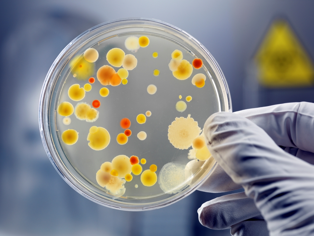
Cultures of “petrophiles”, wild fungi isolated from petroleum contaminated soils in the Ecuadorian Amazon

The other unique aspect of AMP’s approach is the organization’s integration with local communities. Mia Maltz, a fungal ecologist and PhD student at the University of California, Irvine, has been working with the AMP since 2007. Over the last seven years Mia and other members of the AMP have taught mushroom cultivation and mycoremediation (bioremediation using fungi) techniques to locals. By designing and teaching low-cost bioremediation strategies, AMP is empowering locals to do bioremediation on their own.

 “A lot of what we’re doing is very low-tech, easy for local people to do” says Mia. “That’s really the goal here. We want things to be inexpensive and cost-effective. The science here is very simple-  encouraging the natural process of lignocellulose decomposition. We want to show people the simplicity of what’s happening, help them internalize it, so they can tap into a process that’s a huge part of the web of life.”

This summer, a team of scientists and volunteers will work in Sucumbíos to identify and cultivate microorganisms capable of degrading different hydrocarbons present in petroleum. In collaboration with scientists in the United States, the AMP hopes to use metagenomic analyses to profile entire microbial communities and document key microbial taxa and genes involved in petroleum degradation.

AMP volunteer searching for petro-tolerant fungi in contaminated sites

AMP volunteer searching for petro-tolerant fungi in contaminated sites

Ultimately, AMP aims to develop inexpensive biofiltration systems. Biofiltration is the process of filtering contaminated water through living organisms (microbes and plants) which detoxify and remove pollutants. The AMP envisions a biofiltration system consisting of several chambers containing different assemblages of petro-degrading bacteria and fungi. After microbial filtration, the wastewater will be fed to plants that can tolerate high concentrations of heavy metals and other residual contaminants.

AMP volunteers are currently developing prototype biofiltration systems. “We want these to be modular and flexible” says Mia. “We plan on licensing everything in the creative commons to make our technology accessible to local communities around the world.”

The Amazon Mycorenewal Project brings together a broad range of expertise to tackle this enormous pollution challenge. Co-founder Bob Rawson is president of two bioremediation companies (International Wastewater Solutions and the Pseudonym Corporation) and has over 36 years of experience cleaning up contaminated soils. Joanna Zlotnik, program director for AMP, has worked as an environmental geologist for ten years, conducting site assessments and doing remediation work. Collaborator Tradd Cotter is the founder and director of Mushroom Mountain LLC. Tradd brings over 22 years of experience in commercial and experimental mushroom cultivation and mycoremediation research.

Check out the Amazon Mycorenewal Project indiegogo campaign and make a contribution here.

Powering our future with electrigenic bacteria

Standard

Trillions of microbes live on us, in us, and around us, quietly sharing our bodies and our cities. But in the future, some microbes may have to work for their real estate. In fact, they may quite literally become the generators that power our lives. How? By putting them to work inside electricity-generating fuel cells.

The concept of using microbes to generate electricity, otherwise known as a microbial fuel cell, has been around for decades. In essence, a microbial fuel cell is a system that converts chemical energy into electrical energy by taking advantage of the natural oxidation (i.e., electron-release) that occurs as microbes digest organic matter. In this fuel cell, electrons released from microbial digestion are transferred to an electrode. As electrons travel along a charge gradient, they pass through an external electrical connection that harvests some of their energy in a battery or resistor.

Schematic of a microbial fuel cell, in which bacteria connected to an anode digest organic matter, and release electrons that travel to a cathode, generating electric current.

Schematic of a microbial fuel cell, in which bacteria connected to an anode digest organic matter, and release electrons that travel to a cathode, generating electric current. Credit: The Biodesign Institute, Arizona State University.

Scientists have envisioned many ways in which microbial electricity may one day power our lives, from small household electronics to automobiles to self feeding robots. The reality of the matter, however, is that the technology is not yet developed enough to produce substantial quantities of power in a cost-effective way. Most fuel cells today are so large that they can’t fit inside the electronics they are intended to power.

In working to improve microbial fuel cell technology, a lot of effort has been focused on electrochemical engineering. Many scientists are working to improve the efficiency of the electrode: making it better at grabbing and transferring electrons. However, equally important to the development of effective microbial fuel cells is understanding the microorganisms that power them.

In this aspect, one discovery in particular has given scientists hope: electrigens. These are organisms that can harvest energy by directly growing on electrodes. (This is in contrast to the many other organisms that have been scouted for fuel cell application, most of which are simply going about their business digesting organic matter, unaware that some of their precious electrons are being siphoned away to a battery). The most well-studied of the electrigens is Geobacter, an iron-breathing organism that lives in oxygen-free environments. Several species of Geobacter use electrically conductive pili (small antenna-like appendages) to transfer electrons from organic matter directly to iron oxides in the environment. In essence, the process by which Geobacter acquires energy can be co-opted for electricity generation by replacing iron oxides, which occur naturally in soils, with an electrode.

Geobacter coating iron oxide minerals. Credit: Wikipedia

Geobacter coating iron oxide minerals. Credit: Wikipedia

Electrigens represent a promising step towards the development of a sustainable fuel cell, one that can generate a high enough power outputs to be useful. That these bugs naturally thrive on electrodes cuts out one of the largest hurdles associated with microbial fuel cell development. One of the earliest examples of an electrigen-powered fuel cell is the Benthic Unattended Generator (BUG). BUGs live at the bottom of the ocean, producing electric current from organic matter. Their design includes a piece of graphite buried in oxygen-free sediments which serves as an anode. Electrons are collected on this anode when microbes break down sedimentary organic matter. Electrons are transferred to another piece of graphite (the cathode) sitting in the overlying water.

A sediment microbial fuel cell. Credit: Lovely 2006, Nature.

A sediment microbial fuel cell. Credit: Lovely 2006, Nature.

BUGs may one day be used to power monitoring devices and other electronic equipment at the bottom of the ocean. Using a similar concept, scientists have speculated on the possibility of generating electricity from oil spill remediation. Microbes munching away on oil liberate oodles of electrons. Perhaps some of these electrons could be used to power, say, the equipment needed for oil spill remediation.

My personal favorite future use of a fuel cell? Compost generators. Imagine, if you will, an in-home compost bucket that doubles as a generator, allowing you to literally extract power from your trash. That’s a future I’m ready for.

ResearchBlogging.org

Lovley, D. (2006). Bug juice: harvesting electricity with microorganisms Nature Reviews Microbiology, 4 (7), 497-508 DOI: 10.1038/nrmicro1442

Resurrecting ancient microbes to understand evolution

Standard
The ever-growing popularity of the zombie apocalypse is testament to our fascination with the idea of resurrection, or the reanimation of the dead. Credit: Robert D. Brown Inc.

The ever-growing popularity of the zombie apocalypse is testament to our fascination with the idea of resurrection, or the reanimation of the dead. Credit: Robert D. Brown Inc.

When you hear the word “resurrection”, what’s the first thing that comes to mind? Religious miracles? Zombie viruses? The end of the world?

Whatever your mental association, I’m willing to bet it’s not “an emerging scientific discipline.” Well, it just so happens a growing community of microbial ecologists are developing a new use for the traditionally mysticism-affiliated word. These scientists don’t just study modern microbes. Nor do they fashion themselves paleobiologists. No, these people actually straddle the boundary between life and death. They resurrect. And in doing so, they hope to gain insight into how evolution occurs: from single genes to entire communities.

I’ll admit Carl Zimmer beat me to this one , but the topic of resurrection ecology is too interesting and relevant for me to ignore. Zimmer’s article provides an excellent summary of many recent discoveries in the field- including the revival of a 1,500 year old Antarctic moss and the resurrection of a 30,000 year old virus frozen in Siberian permafrost. In previous posts, I’ve discussed both microbial resuscitation from ancient ice  as well as mechanisms some bacteria use to stay alive at subzero temperatures.

Today I’d like to dive a little deeper and really dissect what it is these resurrection ecologists are trying to do. Why resurrect an 8 million year old bacteria, or hatch 700 year old insect larvae? Other than the “cool” factor, is there any profound ecological or evolutionary insight to be gained from this research?

I’ll give you the punchline now: yes, there most certainly is. You see, resurrection of organisms from the past, a feat that was until quite recently considered impossible, offers scientists the opportunity to directly test hypotheses about why evolution occurs.

As an example, imagine a community of bacteria, happily swimming about in a shallow pool of water sitting on top of Siberian permafrost couple thousand years ago. One particularly cold winter, this water freezes and some of these bacteria get trapped in the ice. Perhaps it remains cold enough that the ice doesn’t melt again come spring. Over time, this ice, along with some hapless bacteria, get buried and compacted, frozen in space. But also frozen in time. While some of these guys may  have escaped cryosleep, continuing to grow, reproduce, and die, frozen bacteria don’t change. And because they remain static, evolution also freezes. No reproduction means no gene swapping, no adapting to new environmental pressures, no natural selection. Beyond their ice fortress, genetic cousins of the frozen bacteria are spreading their genes, mutating their DNA. Populations are growing, shrinking and migrating in response to their environment. They are evolving.

Fast forward to the present. Suppose you’re an ecologist studying the bacterial communities of a Siberian lake. You discover that a rare gene known as “cocA” is widespread. What sorts of selective pressures may have caused cocA to become so common? By collecting samples from the surrounding permafrost, you find traces of bacteria frozen in the ice. You extract and sequence their DNA, and determine, to your delight, these frozen organisms are a close genetic match to some of your lake bacteria. Similar, but different enough to infer that some evolution has occurred. Moreover, in all of your frozen samples, a different flavor of the cocA gene- let’s call it “colA” – is dominant.

Here’s where the resurrection part comes in. You’d really like to know how your bacterial communities shifted from colA-dominated to cocA-dominated. To do so, you try growing some of your frozen bacteria in the lab. You give them heat, carbon, and nutrients. Soon, you’ve got fossil bacteria happily multiplying on your lab bench. You have resurrected life.

 More importantly, you’ve got yourself a situation most paleontologists would kill for. You now have living replica of ancient organisms, and their  modern descendants. You set out to conduct a series of lab experiments in which you vary different environmental conditions- pH, nutrient concentrations, temperature- and monitor how your fossil bacteria change. Specifically, you track whether and how your gene of interest (colA) changes over time. What you are really doing is experimentally simulating evolution on an ancient organism. Ultimately, you might be able to determine what sorts of environmental changes must have occurred to lead to the modern, cocA-dominated descendants.

The concept of resurrection ecology is a powerful one. In no other subfield of biology can scientists directly study the ecology or evolution of an organism from a different time. Normally, scientists have to rely on fossils and other “proxies” to make inferences about past species, their ecology, and ultimately, the environment. Imagine what we might learn if we could resurrect a family of dinosaurs and tinker in the lab until we figured out what sorts of environmental conditions lead to the evolution of feathers.

Obviously this is a preposterous idea. Safety considerations aside (we all know how Jurassic Park turned out), dinosaurs reproduce too slowly to observe evolutionary change on useful timescales. Moreover their ranges are too large, and their species interactions too complex, to accurately simulate their habitats.

Microbes are a different story. They require only a petri dish to grow. They reproduce in minutes to hours. Changes in the genetic make-up of microbial communities can be observed within days or weeks. Antibiotic and pesticide resistance are powerful testimonial to the swift pace of microbial evolution.

Resurrection ecology thus represents a compelling new tool for understanding how genes, populations and communities evolve. But its significance may not be limited to the past. By informing population models with “biological archives”, scientists may be able to forecast adaption to future environmental changes. In other words, by helping scientists develop a deeper understanding of how evolution works, resurrection ecology may inform our expectations for the future, and allow us to prepare for it.

*******

A quick update: National Geographic’s cover story last week was on reviving extinct species! Check it out here.

ResearchBlogging.org

Orsini, L., Schwenk, K., De Meester, L., Colbourne, J., Pfrender, M., & Weider, L. (2013). The evolutionary time machine: using dormant propagules to forecast how populations can adapt to changing environments Trends in Ecology & Evolution, 28 (5), 274-282 DOI: 10.1016/j.tree.2013.01.009

Fossil viruses preserved in hot spring bacteria

Standard
Viruses, such as the Avian flu virus depicted in this scanning electron microscope image, are neither alive nor quite dead, but may have a lot to tell us about the evolution of life on Earth. Credit: 3DScience.com

Viruses, such as the Avian flu virus depicted in this scanning electron microscope image, are neither alive nor quite dead, but may have a lot to tell us about the evolution of life on Earth. Credit: 3DScience.com

Fossilized microbes have provided scientists many clues about origins of life. By comparison, little attention is given to viruses in the fossil record. Although technically non-living, there is no question these tiny packets of protein-sheathed DNA have shaped the evolution of most life on earth, including humans. But can viral particles, a fractional size of even the smallest bacteria, actually become fossils? A study recently published in the journal Geobiology argues they can. Here, scientists present the first experimental evidence of silicification– the encasement in silicate minerals- of viruses living in hot spring biofilms. Moreover, these viruses can become fossils while still inside their host cells.

Before we get into viral fossilization, let’s back up for a minute and briefly review what a virus is. Viruses are simple beings that strip the notion of life down to a single, fundamental purpose: propagation of genetic code. While sharing this feature with all life on Earth, viruses are not considered living because they lack the means to carry out genetic reproduction on their own. Instead, viruses propagate by injecting their genetic material into a host cell. One inside a host, a virus uses one of two strategies, or “life cycles”, to reproduce. In the lysogenic cycle, viral DNA integrates with host DNA, lying low and allowing itself to be replicated along with the oblivious host cell. In our own chromosomes, many introns, or non-coding pieces of “junk DNA”, are thought to be ancient viral particles that “went to sleep” inside our cells and never woke up. In the more active (and deadly) lytic cycle, the virus co-opts the host cell’s replication machinery, creating a virus factory that eventually spews forth thousands of baby viruses which proceed to infect other cells.

Conceptual model of the two general modes of viral biology: lytic and lysogenic cycles. Credit: allbiologytutors.blogspot.com

Conceptual model of the two general modes of viral biology: lytic and lysogenic cycles. Credit: allbiologytutors.blogspot.com

Which of these cycles a virus chooses depends a lot on the external environment. If conditions are good in the host cell, it may be advantageous to hunker down and stay put. If, on the other hand, the host cell is already sick or dying, the virus may prefer to replicate as much as possible before abandoning ship.

Hot springs host communities of highly specialized extremeophiles. These bugs are not only hyperthermophilic, or super heat-tolerating, but often cope with extremely acid or alkali conditions. One way they do so is by modifying the pH within their cells, making it closer to neutral. Perhaps unsurprisingly, most hot spring viruses follow a lysogenic cycle, enjoying the less-extreme environment offered by their host.

It was in one such strange habitat, the Gumingquan hot spring in the Yunnan province of southern China, that Dr. Brian Jones and colleagues decided to investigate viral silicification. Why study fossilization in a geothermal hot spring? Geothermal waters, heated by subsurface magma from the Earth’s mantle, are often rich in silica and other minerals, such as calcium and iron, that promote fossilization. Moreover, thick, diverse biofilms hosting “virus-like” particles are known to inhabit the Gumingquan hot spring.

For their study, the researchers collected samples of these biofilms and brought them back to the lab. From biofilms containing many different Bacteria and Archaea, the scientists isolated Geobacillus lituanicus, a thermophilic, aerobic (oxygen-breathing) bacteria belonging to the phylum Firmicutes. They also isolated viral particles by liquifying samples of biofilm, separating out and removing all cells, and filtering the remaining liquid through a 0.22 µm filter- large enough to include viruses but small enough to remove most other microbial debris. Spherical, “virus-like” particles were further isolated and purified by infecting G.lituanicus cells with “viral extract”, and growing the infected cells on agar.

Once the researchers had purified both a host and virus-like particles, they set up a rather clever experiment to test for viral fossilization. They concocted a growth media for G. lituanicus containing sodium metasilicate, a silica-rich mineral. After adding G.lituanicus cells and virus-like particles to this media, they incubated the mixture for 22 days. At regular intervals, the scientists took samples and used transmission electron microscopy (TEM) to search for silicified viruses.

Using TEM, the scientists found numerous silica nanoparticles both inside and outside G. lituanicus cells. These circular particles consist of a core roughly 35-60 nanometers (nm) in diameter, surrounded by an outer shell approximately 30 nm thick. While these particles may just be small, silicified fragments of cellular detritus, there is good reason to think many of them are fossil viruses. For one, the fossil “cores” are similar in size and shape to the spherical virus-like particles used in the experiment. The silica nanoparticles had a “near normal”, or bell-curve, size distribution, another hallmark of biology. Finally, some of the silica nanoparticles had tails.

TEM images showing silica nanoparticles inside and outside cells. Note the light colored "virus-like cores" clearly visible in panels c and d, surrounded by a silicious shell. Credit: Peng et al. 2013

TEM images showing silica nanoparticles inside and outside cells. Note the light colored “virus-like cores” clearly visible in panels c and d, surrounded by a silicious shell. Credit: Peng et al. 2013

A few of you might be scratching your heads at this point, thinking something seems amiss. Remember, I told you most hot spring viruses are lysogenic (integrated into host DNA) most of the time. But in order for a virus to fossilize, it must have its own, independent structure. In other words, it must be lytic.

Does the discovery of fossil viruses within host cells negate the idea that hot spring viruses are typically dormant? Not necessarily. Actually, it may offer an explanation for when and why viral dormancy occurs. You see, all cells eventually die due to accumulated environmental stress. When a hot spring bacteria like G. lituanicus dies, its cell wall breaks open, causing hot, mineral-rich geothermal fluids to rush in. Suddenly, any viruses inside are exposed to all sorts of elements- heat, acid, etc.- they can’t necessarily tolerate. By precipitating a silica shell, these viruses may be able to survive long enough to find a new host.

Some of the proteins on a virus’s outer surface contain chemically-reactive molecules, such as carboxyl groups, that may aid in silica encrustation. The host cell itself may inadventently promote viral fossilization. Many alkaliphiles, like those in the alkaline Gumingquan waters, maintain their cellular pH at two or more units lower than the external environment. Geothermal waters that are undersaturated with silica at pH 12 may become supersaturated at pH 9 or 10, causing silicates to precipitate out as solids.

The strong evidence for viral silicification presented in this experiment suggests viruses may be preserved in the fossil record, particularly in rocks associated with alkaline geothermal systems. Some of the features of virus-like fossils noted in this study- distinct cores and shells, fossil “tails”, a concentration of fossil-like particles inside a host cell- may be useful for identifying the footprints of ancient viruses in the wild.

ResearchBlogging.org

Peng, X., Xu, H., Jones, B., Chen, S., & Zhou, H. (2013). Silicified virus-like nanoparticles in an extreme thermal environment: implications for the preservation of viruses in the geological record Geobiology DOI: 10.1111/gbi.12052

Arctic bacteria gene swap to detoxify mercury

Standard
Mercury, also known as quicksilver, is unusual among metals for being liquid or gaseous at room temperature. This also makes it extremely dangerous for most living organisms. Credit: iStock

Mercury, also known as quicksilver, is unusual among metals for being liquid or gaseous at room temperature. This also makes it extremely dangerous for most living organisms. Credit: iStock

On the list of substances you really don’t want to mess with, elemental mercury is pretty high up. Inhalation or absorption of the volatile metal can quickly lead to irreversible poisoning. Unfortunately, Hg is also a hallmark of industrial society. Fossil fuel combustion releases small amounts of gaseous, elemental mercury, or Hg0,into the atmosphere. Problem is, it usually doesn’t stay that way for long. Free radicals, such as hydroxyl, strip electrons off elemental mercury, converting the metal into its oxidized form, Hg2+

With two electrons missing, mercury falls out of the atmosphere, entering soils and waterways where it can travel readily into plants, animals and people, hungry to restore its charge balance. It turns out oxidized mercury has a high affinity for sulfur. Sulfur, while not abundant in our bodies like carbon or hydrogen, plays an essential role in protein structure. It forms bridges and cross-links, folding our proteins into the shapes needed to carry out their functions. Once in your bloodstream, mercury can scavenge the sulfur out of your proteins, causing them to unravel.

While most higher life forms have no defenses against the devastating effects of mercury poisoning, bacteria have evolved mechanisms to fight the deadly metal. Mercury-resistant bacteria carry a collection of Hg-detoxifying genes, including the enzyme mercuric reductase, which converts Hg2+ back into gaseous H0. How abundant are these guys, and how did mercury resistance evolve? These questions are important for understanding the natural detoxification of mercury-contaminated environments.

In a study recently published in the journal FEMS Microbiology Ecology, Dr. Annette Møller and colleagues seek answers from an unlikely place: the ice sheets and cold, briny waters of the high arctic. Why study mercury detoxifiers in the Arctic? Mercury deposition at the north pole has been increasing for decades due to mercury-enriched air currents making their way poleward from industrializing Asia. Roughly 300 tons of mercury are now deposited across the Arctic every year.

Industrial activity has greatly increased the amount of mobile mercury cycling through the biosphere.

Industrial activity has greatly increased the amount of mobile mercury cycling through the biosphere.

For their study, Møller and colleagues collected snow, fresh water and arctic sea-ice brine from Station Nord in Northeastern Greenland. To isolate mercury resisters, they grew bacteria in media containing low concentrations of mercuric chloride. Once grown, the researchers extracted DNA from their cultures. From these DNA extracts, they determined the identity of mercury detoxifiers by sequencing the 16S ribosomal RNA gene (a marker used to identify different bacterial taxa) . They also sequenced merA, the gene encoding mercuric reductase. By examining the similarity between merA genes from different bacteria, scientists can study the evolutionary history of mercury detoxification and understand how it is transferred through the environment.

In total, the researchers were able to isolate 72 different mercury-resistant bacteria, roughly 10% of the total culturable bacteria. To their surprise, the scientists only found the merA gene in 32% of their mercury-resistant isolates. This may suggest the presence of novel merA genes that are very different from the ones we already know. It could also mean arctic bacteria have different, entirely novel mechanisms of detoxifying mercury.

What of the merA genes the scientists found? merA, it turns out, is diverse, both in its genetic code and taxonomic distribution. Many distantly related bacteria posses the ability to detoxify mercury. Upon further investigation, the scientists discovered that two-thirds of merA carriers also contain plasmids– tiny circular bits of “mobile” DNA that are physically separate from the bacterium’s main chromosome. Plasmids are important agents of horizontal gene transfer, the release of tiny bits of DNA into the environment, where they can be taken up and used by others. The diverse distribution of merA, coupled with the presence of merA plasmids in some bacteria, suggests mercury-resistance may jump through microbial communities, bypassing reproduction and species boundaries.

Plasmids, small DNA molecules physically separate from a bacteria's main chromosome, can be used to transfer DNA between different microorganisms. Credit: Wkipedia commons

Plasmids, small DNA molecules physically separate from a bacteria’s main chromosome, can be used to transfer DNA between different microorganisms. Credit: Wikipedia commons

Thus, arctic microbial communities may adapt to increased mercury concentrations by swapping detoxification genes: much in the same way a piece of code can be shared to patch computer software. This is promising news for folks interested in bioremediation. By releasing mercury detoxifiers in contaminated sites, we may be able to spread the “genetic knowledge”, powering up local microbial communities to combat mercury.

ResearchBlogging.org

Møller, A., Barkay, T., Hansen, M., Norman, A., Hansen, L., Sørensen, S., Boyd, E., & Kroer, N. (2014). Mercuric reductase genes and mercury resistance plasmids in High Arctic snow, freshwater and sea-ice brine
FEMS Microbiology Ecology, 87 (1), 52-63 DOI: 10.1111/1574-6941.12189

When the going gets dense: microbial life under extreme pressure

Standard

From project deadlines to taxes to balancing work and family life, pressure is something we’re all familiar with. Many of us feel like we’re constantly living under pressure. But not like microbes. Microbes take the notion of “living under pressure” to another level entirely.

Life in the deep, dark ocean is mysterious and beautiful. It's also adapted to survive at pressures hundreds to thousands of times greater than humans can tolerate. Credit: National Geographic

Life in the deep, dark ocean is mysterious and beautiful. It’s also adapted to survive at pressures hundreds to thousands of times greater than humans can tolerate. Credit: National Geographic

As I’ve discussed in previous posts, energy limitation and sometimes temperature are two of the main factors restricting microbial life. However, in the deep ocean, the most unique environmental challenge microbes face may be the sheer weight of water bearing down on them.

Welcome to the piezosphere, defined as the part of the ocean where pressure exceeds 10 megapascals (MPa), a hundred times greater than air pressure at sea level. While the piezosphere may seem remote, it is by no means insignificant. It encompasses roughly 75% of the ocean’s total volume, making it the largest habitat for microbial life on earth.

Graphical representation of the piezosphere, where temperature, light and food are scarce, and pressure is high. Credit: Picard and Daniel 2013

Graphical representation of the piezosphere, where temperature, light and food are scarce, and pressure is high. Credit: Picard and Daniel 2013

 Some like it hot, some like it cold

The inhabitants of the piezosphere, aptly named piezophiles, are sub-classified according to the pressure range they tolerate.  The most extreme hyperpiezophiles can tolerate pressures up to 130 MPa, or 1,300 x atmospheric! In the deep ocean, temperatures range from near freezing (2-3 ºC) to piping hot (> 400 ºC near hydrothermal vents). Piezophiles occupy almost the entire spectra of temperatures known to support life (with the exception of some very cold loving bacteria buried in ice and permafrost). Accordingly, scientists further classify piezophiles based on their temperature preference.

All known cold-loving “psychro-piezophiles” are Bacteria, most from the phyla Gammaproteobacteria. These guys thrive in the the deepest, darkest, coldest parts of the ocean and include the most pressure-tolerant organisms on Earth. Species of Shewanella and Moritella, isolated from Challenger Deep, the ocean’s deepest location in the Marianas trench, thrive at pressures of 110 MPa and temperatures of 2 ºC. Other cold-seep bacteria include members of the genus Carnobacterium.  Recently discovered in the Aleutian Trench, these bacteria are close relatives of pyschrophiles found abundantly in Alaskan permafrost.

The deepest part of the ocean floor may not look like much, but it's home to some of the strangest bacteria known to biology. Credit: National Geographic

The deepest part of the ocean floor may not look like much, but it’s home to some of the strangest bacteria known to science. Credit: National Geographic

On the other end of the spectrum, some piezophiles like it hot. Heat and pressure loving microbes, or hyperthermo-hyperpiezophiles  (try saying that five times fast) are more diverse than their cold-weather counterparts, distributed across both Bacteria and Archaea. The hyper-hypers are found near deep sea hydrothermal vents, where fissures in the crust spew hot magma onto the ocean floor. This group includes Methanopyrus kandleri, the most thermophilic (heat loving) bacteria every described, capable of growing at 40 MPa and a whopping 122ºC.

Specialized membranes

How do piezophiles survive under pressures that would be deadly for most life? To answer that question, it’s important to first  understand why high pressure is deadly for most of us. It has to do with our cell membranes. For membranes to function properly, they must remain fluid. Too solid, and many biological processes are interrupted, including cell respiration, nutrient transport and signaling. Low temperatures and high pressures both cause membranes to solidify by “packing”  their building blocks rigidly together.

There is growing evidence that pressure lovers have highly specialized cell membranes. Remember, membranes are composed of fatty acids, molecules with a hydrophilic (water-loving) head and a hydrophobic tail. Fatty acid “tails” are really of a chain of carbon atoms that can vary in length and bonding structure. Many cell membrane properties can be deduced by examining the length and structure of these fatty acid tails.

Several recent studies find piezophile membranes contain unusually high levels of unsaturated fatty acids, or UFAs. UFAs distinguish themselves from saturated fatty acids by their carbon-carbon double bonds, which cause their tails to take on a  kinked structure rather than standing straight. Ultimately, these kinks prevent UFAs from packing too closely together. Hence, more UFAs = more fluid membrane. Piezophile UFAs are unusually long, an adaptation also thought to help maintain their fluidity.

Piezophile membranes are different from regular membranes in that they maintain their structure at extremely high pressures. Credit: Abe 2013

Piezophile membranes are different from regular membranes in that they maintain their structure at extremely high pressures. Credit: Abe 2013

Life Strategies

Pressure aside, life in the piezosphere is not for the faint of heart. Often too cold, sometimes too hot, never enough food. Organic matter enters the deep ocean mainly as intermittent “rain” from the euphotic zone (the sunlit ocean) above. Microbes living off this organic rain are very efficient scavengers, but nonetheless are incredibly slow-growing, suggesting they live in a state of perpetual starvation.

Many organisms in the piezosphere have abandoned carbon entirely, instead eating inorganic compounds like hydrogen sulfide. Some adventurous archaea even make their own food, turning carbon dioxide into sugar just like plants do, but using mineral energy instead of light.

How does pressure affect the diverse metabolism of the piezosphere? The jury’s still out on this one. Generally, in the presence of oxygen, high pressure seems to stimulate deep ocean microbial activity. In the absence of oxygen, the effects of pressure can vary. Iron-reducers (organisms that “breathe iron” instead of oxygen) appear to grow faster at higher pressures, but for other anaerobic (non-oxygen breathing) bacteria, such as sulfate reducers, high pressure appears detrimental.

The high pressure frontier

There are many open questions for scientists interested in the limits of life under extreme pressure. The deep ocean is vast and difficult to sample; its’ microbial diversity is largely unknown. Most sampling efforts have focused on cold seeps and hydrothermal vents. Only a handful of studies examine piezophiles living at more moderate temperatures. Moreover, metagenomic tools, now widely used to examine the diversity of microbial communities, are biased towards abundant individuals. It’s possible that our current genetic tools only capture a fraction of the diversity present in the piezosphere, masking rare but potentially ecologically important players.

Studying the ecology of piezophiles in their “natural environment”  is also a challenge. Specialized equipment can be used to reproduce the high pressures of the piezosphere, but these tools do not necessarily mimic other aspects of the native environment, such as nutrient availability. Most often, researchers are forced to decompress their samples and grow piezophiles under a normal atmosphere. Whether the activity of piezophiles under artificially low pressure is comparable to life in the deep ocean remains to be seen.

All in all,  life in the piezosphere is still shrouded in mystery. Many scientists believe life originated in high pressure environments such as hydrothermal vents or deep ocean trenches. Understanding life in the piezosphere today might help scientists to determine how life on Earth got its start – and, in turn, how life may evolve on other worlds.

ResearchBlogging.org

Picard A, & Daniel I (2013). Pressure as an environmental parameter for microbial life–a review. Biophysical chemistry, 183, 30-41 PMID: 23891571

Fang, J., Zhang, L., & Bazylinski, D. (2010). Deep-sea piezosphere and piezophiles: geomicrobiology and biogeochemistry Trends in Microbiology, 18 (9), 413-422 DOI: 10.1016/j.tim.2010.06.006

Abe, F. (2013). Dynamic structural changes in microbial membranes in response to high hydrostatic pressure analyzed using time-resolved fluorescence anisotropy measurement Biophysical Chemistry, 183, 3-8 DOI: 10.1016/j.bpc.2013.05.005

Deep but not dead: continental crust bacteria make a global impact

Standard
Scanning electron image of mineralized bacteria from a "black smoker", a deep sea hydrothermal vent. Microbes living in extreme subterranean environments such as this may be more abundant than once thought.

Scanning electron image of mineralized bacteria from a deep sea hydrothermal vent known as a “black smoker”. Microbes living in extreme subterranean environments such as this may be more abundant than once thought. Credit : SEM Images

Microbes in the deep biosphere – ocean floor sediments, subterranean aquifers, the continental crust – often get the short end of the research stick. Reproducing every few thousand years and living in perpetual starvation, life far beneath the surface beats to a different drum. The strangeness of life down deep presents numerous challenges to researchers. Getting uncontaminated samples is difficult and expensive, but once they’re back in the lab, inducing “zombie bacteria” to grow is another hurdle entirely.

Given the trouble involved, is it even worth studying the deep biosphere?

Perhaps a different way of framing that question is to ask how much the deep biosphere matters to the rest of the world. Is it simply a collection of curious survivalists, biologically impressive but of little importance to life on the surface? Or is the deep biosphere large enough that we can’t just shrug it off as too remote?

In a paper recently published in the journal FEMS Microbiology Ecology, Dr. Sean McMahon seeks to answer this question using a simple approach. How much, McMahon asks, does the deep continental biosphere weigh?

Ecosystem-scale biomass (or carbon) measurements are used to inform everything from agricultural and land management decisions to climate negotiations. But getting the numbers right is not a trivial problem. A certain amount of assuming, averaging, and extrapolating is always needed. While imperfect, such numbers carry immense value. Estimating the carbon stored in the deep continental biosphere allows scientists to evaluate the importance of subterranean bacteria on a global scale.

To determine the microbial carbon stored in the continental crust, McMahon compiled 120 cell density measurements from aquifers located anywhere from 10 meters to 3.6 km down. He also collected information on the volume of groundwater and its distribution throughout the crust, which he used to estimate the total number of cells present in groundwater worldwide.

McMahon’s groundwater data only represented “unattached” cells. Attachment to mineral particles allows bacteria to form protective biofilms and maintain a relatively stable environment. In aquifers, attached bacteria outnumber free-floaters anywhere from 100 to 1,000-fold. So McMahon scaled up his estimate accordingly. Finally, McMahon assumed that each bacterial cell in the subsurface contains 26 femtograms (10^-15 grams!) of carbon. While the carbon content of a bacteria is highly variable, ranging from 10 – 100 fg, McMahon created a low-end estimate for the weight of an average bacteria living in starvation conditions.

Shewanella oneidensis, a bacteria that attaches to iron oxide clays, using iron as an energy source

Shewanella oneidensis, a bacteria that attaches to iron oxide clays, using iron as an energy source

From all this information, McMahon estimates there are 14 – 135 petagrams of microbial carbon in the continental crust. (1 Pg = 10^15 grams). On the low end, this is approximately twice the amount of carbon that enters our atmosphere every year due to fossil fuel burning. On the high end, it’s roughly equivalent to the total annual carbon dioxide respired by vegetation and soils worldwide.

McMahon’s estimate is probably conservative. Most of his data is from aquifers bearing relatively large sand particles that serve as microbial attachment sites. Sediments consisting of smaller, clay-sized particles have larger surface areas and are likely to host more attached bacteria. McMahon also excluded coal and hydrocarbon deposits from his estimates. But these are energy-rich environments where bacteria are probably far more abundant.

Ultimately, by producing an estimate of  microbial carbon stored in the crust, McMahon is able to affirm the deep biosphere’s global significance. Taken together with new research demonstrating the role subsurface microbes play in rock weathering and nutrient cycling, perhaps the larger take home is that we should start paying a little more attention to the bacteria miles beneath our feet.

An excellent overview of life in the deep biosphere.

ResearchBlogging.org

McMahon S, & Parnell J (2014). Weighing the deep continental biosphere. FEMS microbiology ecology, 87 (1), 113-20 PMID: 23991863

Volcano bacteria uses rare earth metals to survive in acid hot springs

Standard
Rare earth elements, highly coveted in the technology industry, are difficult to mine because of their low concentrations in most rocks. Clockwise from top center: praseodymium, cerium, lanthanum, neodymium, samarium, and gadolinium. Credit: Wikipedia

Rare earth elements, highly coveted in the tech industry, are difficult to mine because of their low concentrations in the Earth’s crust. Clockwise from top center: praseodymium, cerium, lanthanum, neodymium, samarium, and gadolinium.
Credit: Wikipedia

Most of us pay little heed to rare earth elements, found in trace quantities throughout Earth’s crust and sporting unpronounceable names like lanthanum and praseodymium. However, REEs are ubiquitous in our lives, a key ingredient in technological wonders such as cell phones, laptop computers, and solar cells. While our anthropocentric bias may lead us to assume we are the only organisms smart enough to take advantage of REEs, not so, according to a study published last week in the journal Environmental Microbiology. New research indicates that at least one bacteria- perhaps many- use REEs to turn methane into fuel. By doing so, they are able to survive in some of the most extreme environments on Earth.

Just west of Naples, Italy, lies the Solfatara crater: a dormant volcano that spews jets of sulfurous steam from fissures in the Earth. In addition to being the mythological home of Vulcan, the Roman god of fire, Solfatara harbors an unusual member of the bacterial phyla Verrumicrobia known as Methylacidophilum fumariolicum . Discovered in 2007, M. fumariolicum has proven astounding in many ways: it thrives in high heat (120-140 ºF), in strong acid (pH < 1), and with nothing to eat except for methanol, a very simple carbon-containing alcohol. But the ability of scientists to study the unique physiology of M. fumariolicum has been limited by the bacteria’s very slow growth in the lab.

Also known as fumaroles, openings in the planet's crust such as this one emit steam containing a mixture of noxious gases. They are also home to extremeophilic microbes that can tolerate both high heat and acid. Credit: Wikipedia

Also known as fumaroles, openings in the planet’s crust such as this one emit steam containing a mixture of noxious gases. They are also home to extremeophilic microbes that can tolerate both high heat and acid.
Credit: Wikipedia

Until, that is, Dr. Arjan Pol and colleagues from Radboud University decided to add a dash of volcanic mudpot water, M.fumariolicum’s natural habitat, to the petri dish. And, eureka! M.fumariolicum started to grow, at a rate directly proportional to the amount of mud water added.

But this was puzzling to the researchers. They couldn’t find a single element with known biological importance that could replace the mudpot water. In order to narrow down what was causing M.fumariolicum’s growth, the scientists heated the mudpot water to 550ºC to ash off all organic materials. Adding the ashed residue to their cell cultures still caused M.fumariolicum to grow. This clued Dr. Pol in that whatever ingredient M.fumariolicum needed to grow was capable of withstanding high heat- a mineral.

To determine the identity this essential mineral, the scientists further analyzed the mudpot water using inductive coupled plasma mass spectrometry, a technique that allows quantification of different element concentrations. As it turns out, several rare earth elements known as lanthanides were ten times more abundant than usual. By experimentally adding each of these metals to their cultures, the researchers quickly confirmed lanthanides were causing M.fumariolicum to multiply.

Lanthanides are sometimes observed to have positive effects on plant growth, and are commonly used in fertilizers. Scientists speculate that lanthanides boost growth by interacting with Rubisco, the ubiquitous photosynthesis enzyme that binds carbon dioxide and begins the process of turning it into sugar. Metals are often associated with enzymes as cofactors, switches that turn an enzyme on or off by binding or releasing. If a plant’s Rubisco operates more efficiently in the presence of lanthanides, that would mean the plant could turn carbon into sugar faster.

In an analogous manner, Dr. Pol and colleagues guessed lanthanides might be interacting with some protein involved in helping M.fumariolicum acquire energy. To investigate this hypothesis, they extracted and separated different proteins present in their microbe. Lanthanides were most abundant in association with methanol dehydrogenase (MDH), an enzyme that helps break methanol down into methane. Thus it appears this volcanic bacteria requires lanthanide metals in order to metabolize methane, its primary food source.

But why would a bacteria depend on rare earth metals for a crucial component of its metabolism? Most organisms would use more common elements like calcium, zinc or magnesium. It turns out MDH produced by M.fumariolicum has an extraordinarily high affinity for its substrate, methanol. Lanthanides, by slightly altering the enzyme’s 3-D structure, may be responsible for this high affinity. In resource-limited volcanic mud pots, any adaptations that increase an organism’s ability to acquire energy would provide an enormous advantage.

While this study provides the first direct evidence of an organism requiring REEs to grow, use of REEs may not be limited to extreme environments. Certain plants are known to hyperaccumulate REEs in their leaves. Bacteria living on the surface of REE-enriched leaves may be adapted to use these metals. Beaches or ocean sediments are another place for REE lovers might thrive- sand represents a virtually inexhaustible source of these metals.

It's not always easy getting microbes to grow. We can only culture <1% of the bacteria present earth. Bacteria that live in environments very different from our own are poorly represented in the catalog of culturable organisms.

It’s not always easy getting microbes to grow. Currently, scientsts can only culture <1% of the bacteria present earth. Bacteria that live in environments very different from our own are poorly represented in the catalog of culturable organisms.

A major challenge for scientists studying unusual or uncommon microorganisms is growing these critters in the lab. This is often because we are unaware of key ingredients they require. The finding that REEs are essential for some microbes will undoubtedly help future scientists to grow and study more of our planet’s vast unseen diversity.


ResearchBlogging.org

Arjan Pol,, Thomas R. M. Barends,, Andreas Dietl,, Ahmad F. Khadem,, Jelle Eygensteyn,, Mike S. M. Jetten, & Huub J. M. Op den Camp (2014). Rare earth metals are essential for methanotrophic life in volcanic mudpots Environmental Microbiology, 16 (1), 255-264 DOI: 10.1111/1462-2920.12249

Frozen bacteria repair their DNA at -15ºC

Standard

Bacteria encased in ice can be resuscitated after thousands, perhaps even millions of years. How these hardy bugs manage to survive deep freeze is something of a mystery.  If nothing else, the low levels of radiation hitting Earth’s surface should cause any ice-bound bacterium’s DNA to break apart over time, eventually leading to irreparable damage. Some scientists think bacteria survive cryosleep by encasing their DNA in protective shells known as spores and entering a state of dormancy. Following spore formation, a bacterium can withstand harsh environmental conditions, including desiccation, strong acids, heat and UV radiation.

Spores don't actually have eyes like they do in the popular video game, but they can resist drought, fire, ice, acid, and even antibiotics.

Spores don’t actually have eyes like they do in the popular video game, but they can resist drought, fire, ice, acid, and even antibiotics.

But other researchers think we aren’t giving enough credit to the ice dwellers. Recent studies have shown that some psycrhophiles– technical-speak for cold-loving bacteria – are able to maintain basic metabolic functions at subzero temperatures. Could psychrophiles trapped in ice be repairing their DNA faster than the UV radiation bombarding our planet pulls it apart? Microbiologist Markus Dieser at Lousiana State University was interested in finding out. In a study published  in the journal Applied and Environmental Microbiology, Dieser and colleagues show for the first time that one bacteria- Psychrobacter arcticus– can repair it’s DNA at temperatures as low as -15ºC, or 5ºF. Moreover, it can do so 100,000 times faster than damage occurs.

P. articus is an innocuous little bacteria that is famous for one thing: it really likes the cold. It can grow and metabolize at -10 ºC, making it one of the most psychrophilic organisms on Earth. To investigate P. articus’’s ability to repair DNA in deep freeze, Dieser and colleagues isolated viable P. articus cells from Siberian permafrost that has been frozen for 20 to 30 thousand years. In the lab, the researchers dosed their cell cultures with a large pulse of ionizing radiation- roughly equal to what P. articus might experience over 225 thousand years of field exposure. By using such an intense burst of radiation, the team hoped to induce many “double-strand breaks”, or breaks that cause small DNA fragments to separate off from P. articus’s main chromosome.They incubated the irradiated cultures at -15ºC and monitored their survival over the course of 505 days.

Rather astoundingly, the scientists found no significant difference between the survival rates of irradiated and non-irradiated bacteria over the year and a half long study. While this finding alone suggests P. articus can repair its DNA at subzero temperatures, Dieser and colleagues wanted direct evidence.  They used pulse-field electrophoresis, a technique which separates DNA fragments by size, to determine how may DNA double-strand breaks occurred after radiation exposure, and whether the DNA fragments reassembled themselves over time. Like Humpty Dumpty rebuilding himself, the scientists could literally watch P. articus reassemble its genome. On average, P. articus was able to patch thirteen double-strand DNA breaks over the course of the study-  quite close to the roughly sixteen breaks inducted by radiation.

DNA ligase repairing a DNA molecule that has suffered a double-strand break. Credit: Wikipedia

DNA ligase repairing a DNA molecule that has suffered a double-strand break. Credit: Wikipedia

Not only can P. articus repair its DNA at subzero temperatures, it can do so really fast. Using annual radiation exposure data collected in the field, Dieser estimates that P. articus can repair double-strand breaks 100,000 times faster than they occur. The discovery has important implications for the survival of life in extreme environments, including cold extraterrestrial environments. For instance on the surface of Mars, where radiation levels are ~400 times greater than the Siberian permafrost, P. articus can still patch DNA breaks 280 times faster than they would accrue. As scientists continue exploring the “cold limit” to essential cellular functions such as DNA repair, they will continue to refine, and perhaps expand, our understanding of the fundamental boundaries for life.

ResearchBlogging.org

Markus Dieser, John R. Battista, & Brent C. Christner (2013). DNA Double-Strand Break Repair at −15°C Applied and Environmental Microbiology DOI: 10.1128/AEM.02845-13

30,000 feet up, these bacteria aren’t afraid of heights

Standard


The troposphere, the lowermost portion of Earth's atmosphere, is anywhere from 4.3 to 12 miles deep, depending on latitutde.

The troposphere, the lowermost portion of Earth’s atmosphere, is anywhere from 4.3 to 12 miles deep, depending on latitutde.

From ocean floors to office desks, bacteria coat nearly every inch of the Earth. Scientists have recently discovered bacteria are present high above the Earth, as well. Ten kilometers up, to be exact, in a region of our atmosphere known as the upper troposphere. In a place where freezing temperatures, high ozone concentrations and intense UV radiation are toxic for most life, to say nothing of the gravity issue, communities of microorganisms make a home. Furthermore, the “microbiome” of the upper troposphere may play an important role in the formation of clouds and precipitation, according to a study published in the Proceedings of the National Academies of Science.

The NASA-led study began in 2010 as part of an extensive field campaign to better understand how tropical storms develop. Microbiologists worked onboard NASA’s “flying science laboratory”, a renovated Douglas DC-8 jetliner, collecting air samples across the continental US and Caribbean during and after tropical hurricanes Earl and Karl. Collecting each “sample” involved vacuum-pumping six cubic meters of air- the volume of a large van – through specialized filters that trap bacteria-sized particles. The microbes collected on the filters were then studied using high-powered microscopes and genomic techniques to determine their abundance, composition and origin.

While not teeming with life, the upper troposphere is hardly a cold, dead wasteland. The scientists estimated roughly 150,000 living microbial cells per cubic meter of air. (For comparison, there can be billions of microbes in a cubic centimeter of soil). The air samples collected inside Earl and Karl had greater numbers of microbes, many of which are “transplants”, swept out of oceans and soils by powerful hurricane winds. Bacteria are roughly a hundred times more abundant than fungi in the upper troposphere, probably due to their smaller size. Weighing a trillionth of a gram means it can take weeks to fall out of the atmosphere.

Clouds- an underappreciated microbial ecosystem

Clouds- an underappreciated microbial ecosystem

After establishing that bacteria live in the upper troposphere, the scientists set out to determine exactly who’s up there. They did so by analyzing bacterial community DNA using a technique called pyrosequencing. The team uncovered several striking patterns. Perhaps unsurprisingly, hurricanes exhibited the most diverse microbiomes. Post-hurricane, however, there remained dramatic differences in community composition as compared to regions that never experienced hurricanes. Tropical storms appear to leave a lasting legacy on the upper troposphere microbiome, due to the influx of new bacteria and “precipitation scavenging”, or raining out, of preexisting bacteria. In spite of these differences, across all air samples most bacteria fell into two broad groups, the Alpha and Betaproteobacteria. Even more remarkably, seventeen distinct taxa, dubbed the “core microbiome” were present in all samples, from the California coast to the central Caribbean.

Why are the seventeen taxa of the core microbiome so persistent in upper troposphere? In all likelihood, a number of unique characteristics promote the survival of these hardy bacteria. For one, they may have adaptations for coping with UV radiation and high concentrations of oxidants (chemicals like ozone and hydrogen peroxide). Food is another big issue. There is evidence the core seventeen may have more lunch options up high, using the limited carbon sources present in cloud water. For example, Afipia, the most common bacteria in all samples, thrives on a simple carbon compound known as dimethyl sulfone (DMSO2). Compounds similar to DMSO2 are common in marine atmospheres. Two of the other core families, Methylobacteriaceae and Oxalobacteraceae, metabolize oxalic acid, another carbon compound abundant in cloud water. Finally, due to their weight and shape, the core seventeen may simply be harder to rain out.

Cloud water contains simple carbon compounds that provide food for atmosphere-dwelling microbes

Microbial life in the upper troposphere is more than just a curious discovery. These microbes may play an important and unexplored role in cloud formation. Tiny suspended aerosols, previously thought to be non-living, serve as “cloud condensation nuclei” – surfaces that water vapor can condense onto. This study suggests living bacteria may account for 20% of the particles serving as cloud condensation nuclei. And where life is involved, things start to get more complicated. What if different bacteria promote cloud condensation at different rates? Certain plant-pathogenic bacteria actively promote ice condensation on leaf tissue, rendering the plant susceptible to infection. Much as gut microbiomes are now believed to play an important role in our health, atmospheric microbiomes may play an underappreciated role in regulating global weather patterns and climate.